Brain Imaging
|
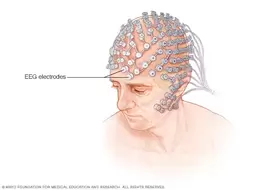
1. Electroencephalograph(EEG): a machine used to record electrical activity of large portions of the brain. Electrical activity can be measured and the rhythms of the brain differ depending on whether a person is awake, drowsy, or asleep. Used for sleep studies.
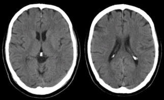
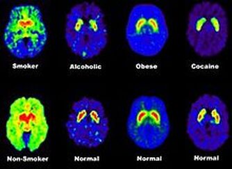
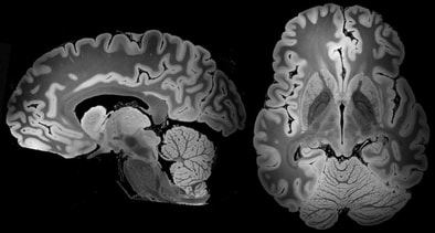
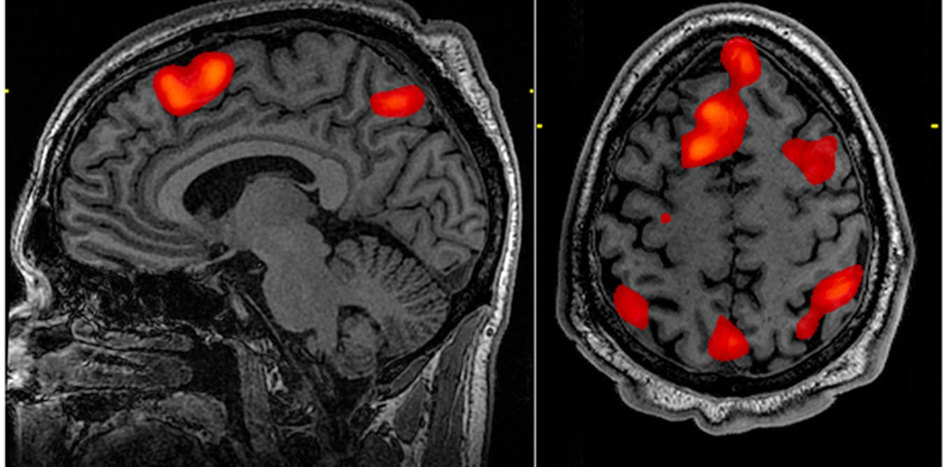
2. Computerized Axial Tomography(CAT/CT): an imaging technique used to study the brain to pinpoint injuries and brain deterioration. Radiation is absorbed depending on the density of the brain tissue. Computers measure the amount of radiation absorbed. 3. Positron Emission Tomography(PET): an imaging technique used to see which brain areas are being activated while performing tasks. Injects radioactive solution into the blood and measures the amount absorbed. Shows activity in different areas of the brain when a person is thinking, speaking, or looking at objects. 4. Magnetic Resonance Imaging(MRI): a measuring technique used to study brain structure and activity, uses no radiation and is excellent for detecting very slight differences in soft tissues. Combines the features of CT and PET scans. 5. Functional Magnetic Resonance Imaging(fMRI): measures brain activity by detecting changes associated with blood flow. Relies on the fact that cerebral blood flow and neuronal activation are coupled. When an area in the brain is in use, blood flow in that region increases. |
|
